Tag: medical history

  • The Wooden Tube That Changed Medicine: A History of the Stethoscope

    The Wooden Tube That Changed Medicine: A History of the Stethoscope

    In 1816, a young French doctor named René Laënnec faced an awkward problem. A female patient with heart trouble was too overweight for him to press his ear directly against her chest the standard method of listening to the lungs and heart at the time. Embarrassed and frustrated, Laënnec remembered a schoolyard trick: sound travels through solid objects. He rolled a quire of paper into a tight cylinder, placed one end on her chest, and put his ear to the other. To his astonishment, he heard her heart sounds “much more clearly and distinctly” than ever before.

    That rolled paper was the first stethoscope, an invention that would transform medicine from an art of guesswork into a science of listening. Yet while Laënnec is often credited as the sole inventor, the stethoscope’s story is actually a chain of forgotten contributors—from Hippocrates’s ear-on-chest listening to the modern acoustic design by David Littmann. Each step brought the instrument closer to what we now take for granted: a doctor’s ability to hear inside the body.

    Before the Stethoscope: Listening with Bare Ears

    For centuries, physicians had only their senses to diagnose illness. They would ask patients about symptoms, look at their bodies, feel for lumps, and—when they needed to hear the heart or lungs—press an ear directly against the chest. This is called immediate auscultation, from the Latin for “listening” (auscultare). It was a practice that dated back to Hippocrates around 400 BCE, who described hearing a “succession” sound like shaking a half-filled bottle inside the chest of a patient with fluid in the lungs.

    But direct listening had serious drawbacks. It was socially awkward, especially with female patients, who might be modest about exposing their chests. It was unhygienic, as doctors came into contact with skin, sweat, and potential infections. And it was imprecise: sounds from different parts of the lung or heart were hard to distinguish, and an obese patient’s chest wall muffled the sounds altogether.

    The first step toward solving this came from an unlikely source: a Viennese physician named Leopold Auenbrugger. In 1761, he published a book describing percussion—tapping the chest with fingers and listening to the resulting sound to determine if organs were solid or filled with air. His method was largely ignored for decades until it was translated and promoted by Jean-Nicolas Corvisart, Napoleon’s personal physician, in 1808. Corvisart was also Laënnec’s mentor, and his advocacy of percussion helped create a culture of systematic physical diagnosis in Paris.

    The Eureka Moment: A Rolled Paper Cylinder

    The story of the stethoscope’s invention is well documented because Laënnec himself described it in his 1819 treatise De l’auscultation médiate. In 1816, at the Hôpital Necker in Paris, he was consulted by a young woman with heart palpitations. She was overweight, and direct auscultation was both impractical and improper. Laënnec recalled a childhood memory: children scratching one end of a wooden beam and hearing the sound clearly at the other end, demonstrating that sound travels efficiently through solid materials.

    He quickly rolled a quire of paper (about 24 sheets) into a tight cylinder, tied it with string, and applied one end to her chest. The result was immediate and dramatic. He could hear the heart sounds with remarkable clarity, much better than with direct ear contact. This was the birth of mediate auscultation—listening through an instrument.

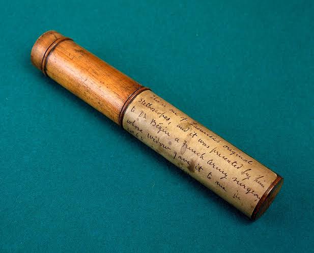
    Laënnec then refined his design into a hollow wooden tube, about 30 centimeters long and 4 centimeters in diameter, with a funnel-shaped chest piece that could be removed. He named it the stethoscope from the Greek stēthos (chest) and skopein (to examine)—literally a “chest examiner.” In 1819, he published his findings in a landmark book that described the sounds of various lung and heart conditions, correlating them with autopsy findings. This was a revolutionary approach: instead of guessing from symptoms, doctors could now hear specific sounds—rales, rhonchi, crepitations—and match them to physical abnormalities.

    The wooden tube was not immediately embraced. Some physicians ridiculed it as a “toy” or a “trumpet,” arguing that it distanced the doctor from the patient. Others found it difficult to use. But Laënnec’s meticulous correlations, his hundreds of autopsies, and the clear benefit of diagnosing tuberculosis (a disease that killed him in 1826) gradually won over the medical community. The treatise was translated into English and German, spreading the technique across Europe.

    The Forgotten Innovators: Who Made the Stethoscope Practical

    While Laënnec deserves credit for the original invention, the stethoscope we use today is not his wooden tube—it is the product of several lesser-known innovators. These are the “forgotten inventors” who each solved a critical problem.

    Pierre Adolphe Piorry and the Pleximeter

    In 1828, French physician Pierre Adolphe Piorry (1794–1879) introduced the pleximeter, a small plate, often made of ivory or wood, placed on the chest during percussion. This made tapping louder and clearer than using fingers alone. Piorry also modified the stethoscope, making it trumpet-shaped with a wider bell to capture more sound. He was a pioneer in correlating percussion and auscultation findings, but his contributions are often overshadowed by Laënnec’s fame.

    Arthur Leared and the Binaural Stethoscope

    The monaural (single-ear) stethoscope forced doctors to lean awkwardly, one ear to the patient’s chest. In 1851, Irish physician Arthur Leared invented a binaural (two-ear) version, but it was rigid and impractical. The following year, American George Cammann (1804–1863) perfected a flexible binaural stethoscope with two ear tubes that could be worn comfortably. Cammann’s design made the stethoscope practical for routine use, allowing doctors to listen through both ears and hear sounds more clearly. Yet Cammann is barely mentioned in standard histories.

    Rappaport and Sprague: The Bell and Diaphragm

    In the 1940s, cardiologists Maurice Rappaport and Howard Sprague designed a stethoscope that combined two chest pieces: a bell (open cup) for low-frequency sounds like heart murmurs, and a diaphragm (flat plate) for high-frequency sounds like breath sounds. This combination allowed doctors to switch between the two by pressing harder or turning the chest piece. The Rappaport-Sprague design became the standard for decades, but it was heavy and had multiple parts that could leak.

    David Littmann: The Modern Acoustic Stethoscope

    In the 1960s, Harvard cardiologist David Littmann (1906–1981) revolutionized stethoscope design. He created a lightweight instrument with a single chest piece that housed both a bell and a diaphragm, using a sealed acoustic system to minimize sound loss. The Littmann stethoscope became the gold standard, known for its clear acoustics and comfort. Today, most medical students receive a Littmann as their first stethoscope.

    The Stethoscope’s Legacy: How It Changed Medicine

    The stethoscope was not just a new gadget; it fundamentally altered how doctors think. Before Laënnec, medicine relied heavily on patient narratives and subjective impressions. The stethoscope enabled clinicopathological correlation—linking specific sounds to specific diseases confirmed at autopsy. This shifted medicine toward objective, evidence-based diagnosis, a cornerstone of modern practice.

    It also made tuberculosis, the “white plague” of the 19th century, more diagnosable. Laënnec himself used his invention to study the disease, and his early diagnoses likely saved lives even if there was no cure. Ironically, Laënnec contracted tuberculosis from his patients and died of it in 1826, at age 45.

    Socially, the stethoscope was initially controversial because it seemed to depersonalize the doctor-patient relationship. Some critics feared it would replace hands-on care. But over time, it became a symbol of medical professionalism—a doctor’s tool that signals expertise and care.

    The Future: Electronic and Digital Stethoscopes

    Since the 1990s, electronic and digital stethoscopes have emerged. They amplify sounds, filter background noise, and can even record and transmit sounds for telemedicine. Some use Bluetooth to connect to apps that display waveforms or analyze sounds with artificial intelligence. Yet despite these advances, the basic principle remains the same as Laënnec’s rolled paper: capturing the subtle vibrations of the body and translating them into diagnostic information.

    The wooden tube may be a relic, but the stethoscope itself endures as one of medicine’s most iconic and essential tools. And its history reminds us that great inventions are rarely the work of a single genius. They are built on the contributions of many—some famous, some forgotten—who each added a piece to the puzzle.

    The stethoscope’s journey from a rolled paper cylinder to the modern acoustic instrument is a story of incremental innovation. Laënnec’s initial spark was brilliant, but it was Piorry, Cammann, Rappaport and Sprague, and Littmann who made the stethoscope practical, comfortable, and effective. As you listen to a doctor’s stethoscope today, remember the forgotten minds that shaped it—and the humble wooden tube that started it all.

    Summary

    • The stethoscope was invented in 1816 by French physician René Laënnec, who used a rolled paper cylinder to listen to a patient’s chest.
    • Before the stethoscope, doctors used direct ear-to-chest listening, which was awkward, unhygienic, and imprecise.
    • Laënnec’s wooden monaural stethoscope enabled mediate auscultation and led to clinicopathological correlation, transforming medicine into an objective science.
    • Key forgotten innovators include Pierre Piorry (pleximeter), George Cammann (binaural stethoscope), Rappaport and Sprague (bell/diaphragm), and David Littmann (modern acoustic design).
    • The stethoscope remains essential, with electronic and digital versions now available, but its core principle has not changed in over 200 years.

    FAQ

    Q: Who really invented the stethoscope?
    A: René Laënnec is credited with the invention in 1816, but many others improved it. George Cammann made it binaural, Rappaport and Sprague added the bell/diaphragm, and David Littmann refined the modern acoustic design.

    Q: Why was the stethoscope initially criticized?
    A: Some physicians thought it was a toy or that it distanced the doctor from the patient. Others found it hard to use. It gained acceptance after Laënnec’s book was translated and its diagnostic benefits became clear.

    Q: What is mediate auscultation?
    A: It means listening to internal body sounds using an instrument, as opposed to immediate auscultation where the ear is placed directly on the body. The stethoscope made mediate auscultation possible.

    Q: How did the stethoscope change medicine?
    A: It allowed doctors to correlate sounds with autopsy findings, leading to clinicopathological correlation and a more objective, evidence-based approach to diagnosis. It also improved diagnosis of conditions like tuberculosis.

    Q: What is the future of the stethoscope?
    A: Electronic and digital stethoscopes can amplify sounds, filter noise, and transmit data for telemedicine. Some use AI to analyze sounds. But the basic principle remains the same as Laënnec’s original invention.
  • The Mold That Changed Medicine: How a Dirty Lab Bench Gave Us Penicillin

    The Mold That Changed Medicine: How a Dirty Lab Bench Gave Us Penicillin

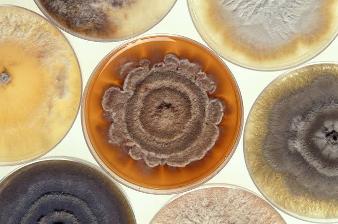
    In September 1928, a Scottish bacteriologist named Alexander Fleming returned from a two-week holiday to find a messy laboratory bench in London. Among a stack of dirty Petri dishes, one had been contaminated by a stray mold. Instead of tossing it out, Fleming took a closer look and noticed something remarkable: the mold had created a clear ring where bacteria had been destroyed. That chance observation set off a chain of events that would eventually save hundreds of millions of lives.

    This is the story of how a fortunate accident, combined with years of painstaking work by many scientists, turned a curious mold into the world’s first antibiotic. It’s a tale of missed opportunities, wartime urgency, and the power of collaborative science.

    A Messy Bench and a Lucky Break

    Fleming was not a tidy researcher. His lab at St. Mary’s Hospital was cluttered, and he often left cultures out for weeks while he attended to other matters. Before leaving for vacation, he had stacked several Petri dishes seeded with Staphylococcus bacteria, intending to clean them later. When he returned, he noticed that one dish had a patch of mold growing on it. Most people would have seen only a ruined experiment. Fleming saw something else: a halo of clear agar around the mold, where the bacteria had been killed.

    He later remarked, “That’s funny,” and instead of discarding the plate, he took a sample of the mold. It was later identified as Penicillium notatum, likely drifted from a mycology lab downstairs where a colleague was studying mold allergens.

    The First Hints of a “Wonder Drug”

    Fleming found that the mold produced a substance that killed many harmful bacteria, including those causing strep throat, pneumonia, and diphtheria. He named it penicillin. He also tested it on animals and found it surprisingly non-toxic. In 1929, he published his findings in the British Journal of Experimental Pathology, but the paper received little attention.

    Why didn’t Fleming pursue it further? The main obstacle was that he could not isolate the active ingredient. The penicillin in the mold broth was unstable, and he lacked the chemistry skills to purify it. He tried using crude filtrates to treat eye infections with some success, but results were inconsistent. By the early 1930s, he had moved on to other research, and the mold sat in storage.

    Enter the Oxford Team

    A decade later, a team at Oxford University picked up the thread. Howard Florey, a pathologist, and Ernst Chain, a biochemist, were interested in natural antibacterial substances. They came across Fleming’s paper and decided to investigate. With the help of biochemist Norman Heatley, they developed methods to extract and purify penicillin from the mold broth. Heatley’s ingenuity was crucial: he designed a freeze-drying process to concentrate the drug and used solvent extraction to purify it. They also devised ways to grow the mold in large quantities using improvised containers like bedpans and milk churns.

    In May 1940, they tested their purified penicillin on mice infected with lethal doses of streptococci. All treated mice survived; all untreated mice died. It was a spectacular result, but they needed to test it on humans.

    The First Human Patient

    In February 1941, they treated their first patient, a policeman named Albert Alexander, who had a severe facial infection that had spread to his eyes and scalp. Penicillin worked wonders—Alexander improved dramatically within a day. But the supply was limited, and the team had to extract penicillin from his urine to reuse it. When the supply ran out, Alexander relapsed and died. It was a heartbreaking setback, but it proved the drug’s power.

    The Oxford team published their results in The Lancet in August 1940, but Britain was at war and could not mass-produce penicillin. They needed help.

    The American Solution

    In mid-1941, Florey and Heatley traveled to the United States to seek industrial partners. They ended up at the USDA’s Northern Regional Research Laboratory in Peoria, Illinois. There, scientists worked on two key problems: finding a better mold strain and developing a method for large-scale production.

    A breakthrough came when a lab assistant brought in a moldy cantaloupe from a local market. The mold on it, Penicillium chrysogenum, produced far more penicillin than the original strain. Another breakthrough was the development of deep-tank fermentation, led by chemical engineer Margaret Hutchinson Rousseau. This method grew the mold in large vats using a nutrient-rich corn steep liquor, allowing production on an industrial scale.

    Pharmaceutical companies like Pfizer, Merck, and Squibb joined the effort, coordinated by the US government. By D-Day in June 1944, there was enough penicillin to treat all Allied casualties. It was hailed as “the wonder drug.”

    The Nobel Prize and the Unsung Hero

    In 1945, Fleming, Florey, and Chain shared the Nobel Prize in Physiology or Medicine. Norman Heatley, whose practical genius made the purification and production possible, was not included—a decision that many historians consider a grave oversight. Heatley’s contribution was essential: without his freeze-drying and extraction techniques, penicillin might have remained a laboratory curiosity.

    The Legacy of Penicillin

    Penicillin’s discovery transformed medicine. It turned once-fatal infections into treatable conditions and sparked the development of other antibiotics. It also highlighted the role of chance in science, but as Louis Pasteur once said, “Chance favors the prepared mind.” Fleming’s curiosity and the Oxford team’s persistence turned an accident into a life-saving breakthrough.

    Today, antibiotics save millions of lives each year, but their overuse has led to antibiotic resistance, a growing threat. The story of penicillin reminds us of the delicate balance between discovery and stewardship.

    The discovery of penicillin is a powerful example of how a chance event, combined with scientific rigor, can change the world. It also underscores the importance of collaboration and the need to preserve the effectiveness of antibiotics for future generations. As we face new challenges like antibiotic resistance, the lessons from that moldy Petri dish remain as relevant as ever.

    Summary

    • In 1928, Alexander Fleming accidentally discovered that a mold called Penicillium notatum killed bacteria, naming the active substance penicillin.
    • Fleming could not purify penicillin, so the project stalled until a team at Oxford (Florey, Chain, and Heatley) developed methods to extract and purify it in the early 1940s.
    • The first human trial in 1941 was promising but ran out of drug, leading to the patient’s death.
    • American scientists and pharmaceutical companies, with the help of a moldy cantaloupe, developed large-scale production methods, making penicillin widely available by 1944.
    • Fleming, Florey, and Chain won the Nobel Prize in 1945, but Heatley’s crucial contributions were overlooked.

    FAQ

    Q: What exactly did Fleming observe on the Petri dish?
    A: He saw a mold colony surrounded by a clear ring where staphylococci bacteria had been destroyed, indicating the mold produced a bacteria-killing substance.

    Q: Why didn’t Fleming develop penicillin himself?
    A: He lacked the chemistry skills to isolate and purify the unstable compound, and his crude extracts gave inconsistent results, so he abandoned the project.

    Q: Who were the key people at Oxford that made penicillin a reality?
    A: Howard Florey, Ernst Chain, and Norman Heatley. Heatley’s techniques for purification and large-scale growth were essential.

    Q: How did American scientists help mass-produce penicillin?
    A: They found a better mold strain from a cantaloupe and developed deep-tank fermentation using corn steep liquor, enabling industrial production.

    Q: Why didn’t Norman Heatley share the Nobel Prize?
    A: The Nobel committee only awarded it to Fleming, Florey, and Chain, but many historians believe Heatley’s contributions were equally deserving of recognition.

  • How the Trenches Forged Modern Medicine: The Medical Transformation of WWI

    How the Trenches Forged Modern Medicine: The Medical Transformation of WWI

    On July 1, 1916, the first day of the Battle of the Somme, British forces suffered over 57,000 casualties—the bloodiest day in British military history. The wounded came in waves, often with limbs torn by shrapnel, faces shattered by machine-gun fire, and bodies contaminated by the filth of the trenches. The existing medical system, designed for an earlier era of warfare, was overwhelmed within hours.

    Yet out of this catastrophe emerged a new kind of medicine. The war forced innovation at a pace peacetime would never have allowed. From the formalization of triage to the birth of plastic surgery, the lessons learned on the Western Front and beyond reshaped how we treat trauma, infection, and psychological injury—and their impact is still felt in every emergency room today.

    The Surgeon Closes In: Mobile Units

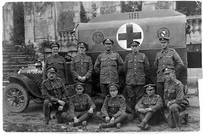
    Surgeons realized that time was the enemy. The French developed the autochir, a mobile surgical hospital that could be moved close to the front lines. The British and Americans followed with casualty clearing stations, which brought surgical teams within a few miles of the trenches. This reduced the time between injury and surgery from days to hours, dramatically improving survival rates for abdominal and chest wounds that had previously been almost always fatal.

    Wound Care: The Debridement Revolution

    The war taught surgeons that antiseptics alone were not enough. The standard practice became debridement—surgically cutting away all dead and contaminated tissue—followed by delayed primary closure. Wounds were left open to drain for several days before being closed. This approach, though painful and labor-intensive, reduced the incidence of gas gangrene and sepsis. It remains a fundamental principle in trauma surgery today.

    Blood Transfusion: From Risk to Routine

    Before WWI, blood transfusions were rare and often fatal due to incompatible blood types. The earlier work of Karl Landsteiner on blood typing, combined with the discovery of sodium citrate as an anticoagulant, made safe indirect transfusions possible. By 1917, the British Expeditionary Force had mobile blood transfusion teams. This innovation meant that soldiers who would have bled to death could be resuscitated and taken to surgery. The principles of blood banking and transfusion established during the war became standard in civilian medicine.

    Faces Rebuilt: The Birth of Plastic Surgery

    Trench warfare produced a devastating new type of injury: facial mutilation. Shrapnel and machine-gun fire tore away jaws, noses, and cheeks. These soldiers survived but faced lives of isolation and horror. New Zealand-born surgeon Harold Gillies established a dedicated hospital at Sidcup, England, where he performed over 11,000 operations. He developed techniques for rebuilding faces using skin grafts and bone transplants, many of which are still in use today. Gillies is considered the father of plastic surgery.

    X-Rays at the Front: Marie Curie’s Petites Curies

    Marie Curie, already famous for her work on radioactivity, recognized that X-rays could help surgeons locate bullets and shrapnel before operating. She developed mobile X-ray units, nicknamed “petites Curies,” which were driven to field hospitals. These units allowed surgeons to operate with precision, removing foreign objects with less tissue damage. Curie herself trained radiologists and drove ambulances to the front, bringing cutting-edge technology to the battlefield.

    The Recognition of Shell Shock

    The term “shell shock” entered medical vocabulary during WWI, describing the psychological collapse of soldiers under prolonged artillery bombardment. Initially, many commanders and doctors dismissed it as cowardice or malingering, fearing that evacuation would encourage others to fake symptoms. But the sheer number of cases—men who trembled uncontrollably, were paralyzed without physical cause, or lost their speech—forced the medical establishment to acknowledge combat-related psychological injury. Early psychiatric treatment programs were flawed, often using electric shock or hypnosis, but they laid the groundwork for modern combat psychiatry.

    Nurses: The Backbone of Care

    Over 20,000 British nurses served overseas, and their role expanded dramatically. Beyond basic care, they administered anesthesia, dressed wounds, and assisted in surgery. The war accelerated the professionalization of nursing, leading to formal training and credentials. Nurses like Edith Cavell, who helped soldiers escape occupied Belgium, became symbols of sacrifice and skill. The war demonstrated that nursing was not just compassion but a highly skilled profession essential to medical care.

    Fighting Disease: Vaccination and Sanitation

    Disease had killed more soldiers than bullets in previous wars. During WWI, mass vaccination campaigns against typhoid and tetanus were implemented. The U.S. Army’s typhoid vaccination program reduced typhoid deaths from 5,000 during the Spanish-American War to fewer than 100 in WWI. Lice control, sanitation measures, and mobile laboratories also helped prevent trench fever and other diseases. These public health measures were as important as surgery in keeping soldiers alive.

    The Great War was a crucible that forged modern medicine. The innovations—triage, debridement, blood transfusion, plastic surgery, mobile X-rays, psychiatric care, professional nursing, and mass vaccination—were born of necessity and refined under fire. They transformed military medicine and, in the decades after the war, seeped into civilian practice, saving millions of lives. The next time you see an emergency room triage team, a trauma surgeon cleaning a wound, or a face transplant, you are seeing the legacy of those who fought and healed in the trenches.

    Summary

    • WWI’s casualty scale forced a dramatic overhaul of military medicine, reducing mortality among the wounded from ~14% (Civil War) to under 8%.
    • Triage was formalized to prioritize surgical resources for the most savable patients.
    • Mobile surgical units and casualty clearing stations shortened the time from injury to surgery, saving lives.
    • Debridement and delayed primary closure became standard wound care, reducing infections.
    • Blood typing and anticoagulant-citrate made safe transfusions possible, leading to mobile transfusion teams.
    • Plastic surgery emerged to rebuild shattered faces, with Harold Gillies pioneering techniques used today.
    • Marie Curie’s mobile X-ray units brought radiology to the front, aiding surgical precision.
    • “Shell shock” forced recognition of psychological trauma, laying groundwork for combat psychiatry.
    • Nursing became professionalized, with women taking on advanced roles.
    • Mass vaccination against typhoid and tetanus dramatically reduced disease deaths.

    FAQ

    Q: What is triage and how did it start?
    A: Triage is the sorting of patients by severity of injury to prioritize treatment. In WWI, French and British armies formalized it, categorizing the wounded as immediate, delayed, expectant, or walking. It became standard in emergency medicine.

    Q: How did blood transfusion become safe during WWI?
    A: Earlier work on blood typing by Karl Landsteiner, combined with sodium citrate as an anticoagulant, allowed blood to be stored and transfused safely. By 1917, mobile transfusion teams deployed near the front.

    Q: What was the role of Marie Curie in WWI?
    A: Marie Curie developed mobile X-ray units, called “petites Curies,” that brought radiology to field hospitals. She also trained radiologists and personally drove ambulances to the front, helping locate bullets and shrapnel in wounded soldiers.

    Q: How did WWI change plastic surgery?
    A: Facial injuries from trench warfare led to pioneering reconstructive surgery. Harold Gillies established a dedicated hospital at Sidcup, performing over 11,000 operations and developing techniques still used in plastic surgery today.

    Q: What was “shell shock” and how was it treated?
    A: Shell shock was a term for psychological trauma caused by the horrors of trench warfare. Initially dismissed as cowardice, the huge number of cases forced recognition of combat-related PTSD. Early treatments were flawed but led to modern psychiatric care for veterans.